National Research Council Canada - National Science Library Post- Processing Resolution Enhancement of Open Skies Photographic Imagery Conclusion RME performed on children in stages of primary and mixed dentition did not have any impact on nasal morphology, as assessed using facial analysis. In 4.92% of the patients between the immediate post-expansion period and 1 year following expansion and in 6.56% of the patients between the pre-expansion period and one year following expansion. Alterations were only detected in the nasolabial angle in 1.64% of the patients between the pre-expansion and immediate post-expansion photographs.

Results From the analysis of the mode of the examiners' findings, no alterations in nasal morphology occurred regarding the following aspects: dorsum of nose, alar base, nasal width of middle third and nasal base. Intraexaminer and interexaminer agreement (assessed using the Kappa statistic) was acceptable. The examiners were instructed to assess nasal morphology and had no knowledge regarding the content of the study. Material and Methods Facial photographs (front view and profile) of 60 patients in the pre-expansion period, immediate post-expansion period and one year following rapid maxillary expansion with a Haas appliance were evaluated on 2 occasions by 3 experienced orthodontists independently, with a 2-week interval between evaluations. Objective The aim of the present study was to use facial analysis to determine the effects of rapid maxillary expansion (RME) on nasal morphology in children in the stages of primary and mixed dentition, with posterior cross-bite. Photographic assessment of nasal morphology following rapid maxillary expansion in childrenĭa SILVA FILHO, Omar Gabriel LARA, Tulio Silva AYUB, Priscila Vaz OHASHI, Amanda Sayuri Cardoso BERTOZ, Francisco Antônio Final reportĬontents: Sensitometers, densitometers, and testing equipment Pitfalls of the photographic (and radiographic) process Evaluation and optimization of photographic processes Quality assurance Odds 'n' ends Photographic processing, quality assurance, and the evaluation of photographic materials. Photographic quality assurance in diagnostic radiology, nuclear medicine, and radiation therapy. Recently obtained PIV photographs are analyzed. The data acquisition and analysis procedure is described, and results of a preliminary simulation study to evaluate the accuracy of the technique are presented. The technique avoids the need for using Fourier transforms with the associated computational burden. Prospects of application of sets of diffusion photographic materials in the practice of rapid X-ray radiography to solve the problems of industrial X-ray defectoscopy are panted outĪ histogram-based technique for rapid vector extraction from PIV photographsĪ new analysis technique, performed totally in the image plane, is proposed which rapidly extracts all available vectors from individual interrogation regions on PIV photographs. Selts of diffusion photographic materials are developed, which contain bromoiodosilver negative material with silver spraying of 2-3 g/m 3 and transparent positive material on Dacron basis.


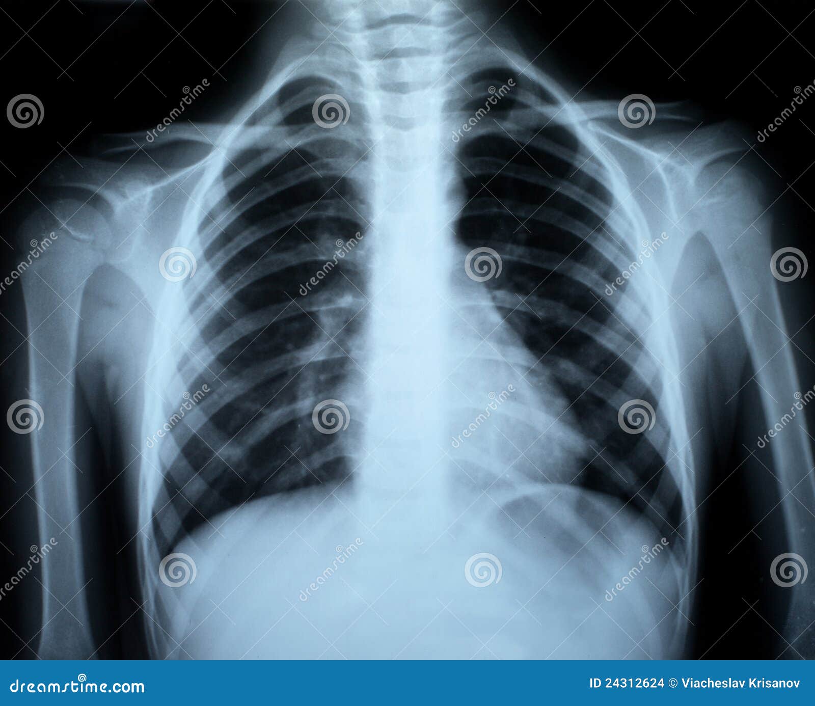
It is shown that the use of diffusion photographic materials for X-ray radiography permits to reduce the process labour consumption and to considerably reduce the time for the obtaining of a dry positive image and also to reduce the consumption of silver, but the image will preserve high information content. Peculiarities of application in X-ray radiography of roentgenographic films, roentgenographic paper, xeroroentgenographic plates and sets of diffusion photographic materials are considered.

International Nuclear Information System (INIS) Characteristics of sets of diffusion photographic materials for rapid X-ray radiography


 0 kommentar(er)
